BIOPHOTONICS
Introduction
nterdisciplinary approaches using the concepts and tools of physics contribute significantly to advances in the understanding of biological mechanisms at the cellular and tissue scales. The exploitation of photon/living matter interactions through microscopy or endoscopy represents major contributions. At PhLAM, this research area is led by the DySCo, Photonics and PMI research teams. The development of advanced microscopy techniques and modelling methods for complex systems is a major focus at PhLAM. These rely in particular on the development of microscopy methods and data analysis tools to quantify interactions, reactions (biosensors) and molecular dynamics in cells, and to model regulatory networks.
The Biophotonics Platform, created in 2016, brings together the biology, microscopy and microfluidics expertise and technical resources of PhLAM in order to enable the development and use of cell biology and photonic microscopy techniques, as well as microfluidics. The platform's mission is to contribute to the design, adaptation and experimental testing of new biophotonics methodologies at the interface between physics and biology. It draws on the interdisciplinary expertise and developments of the members of the Biophysics group within the DYSCO team.
The Biophotonics Platform is open to external users, particularly for expertise not available at other Lille technical platforms (such as fluorescence lifetime imaging microscopy, fluorescence correlation spectroscopy, single particle tracking, Raman spectroscopy, laser optoporation, and cell dynamics measurement).
For more information on the Biophotonics Platform: phlam-biophotoniqueuniv-lille.fr
Activities and skills
- Cell biology
- Molecular biology
- Microscopy
- Microfluidics
- Image analysis
- Modelling
- Biosensor
- Molecular and cellular dynamics
Laurent HELIOT (IR HC)
Technical Director of PhLAM and Research engineer - Biophotonics manager and member of the DYSCO team
Emmanuel COURTADE (MCHC)
Lecturer - DYSCO team leader
Equipment, technologies and achievements
Biology
The biology facilities are located on the 2nd floor of building P5, covering an area of 115 m²: a cell culture room, molecular biology and biochemistry rooms, and a microbiology room. The biological experiments carried out in the laboratory are based on the use of cell lines with genetically encoded markers to track, measure and analyse molecular events in cells. Cell culture is performed in a BSL2 laboratory. Over the past four years, the platform has implemented the CRISPR/Cas9 technique, which represents the state of the art in the field. It enables the direct integration into the genome of the genetic information encoding a fluorescent protein fused to the protein of interest. Building on this technique, BTF has also developed molecular tools to identify the localisation of genes and nascent RNAs (dCas9/dCas13).
Microfluidics
In recent years, we have begun developing microfluidic techniques, building on the expertise of our colleagues at IEMN. Microfluidic techniques can be used to create cellular environments that mimic physiological conditions (i.e. organs-on-a-chip), or combined with microscopy for high-throughput applications to study cellular responses to physiological or therapeutic agonists. A microfluidic technique is also currently being developed for a new photoporation-based transfection system.
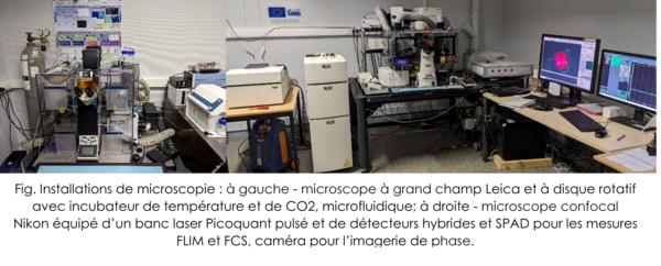
Microscopy
The microscopy facilities, located in the basement of building P5, include: (1) A Leica wide-field microscope equipped with a Spinning-disk confocal and a laser bench with power modulation for phase modulation measurement applications (FLIM). This setup is equipped with a CO2 incubator and temperature control. It is also fitted with an automated perfusion system to induce metabolic changes or stress during multi-day cell monitoring. (2) A Nikon scanning confocal microscope equipped with a temperature and CO2 incubator. Based on a development initiated in the laboratory in collaboration with the company Picoquant, this microscope is equipped for FLIM and FCS measurements. Using these instruments, we develop methods to measure molecular dynamics and track molecular activity in cells over several days using biosensors. (3) A cell photoporation setup coupled to a microfluidic device. (4) A PALM-type super-resolution microscope and single-molecule tracking in living cells in HiLO mode (research team).